Modern dentistry has undergone a massive transformation over the past few decades, moving far beyond basic manual examinations and traditional visual checks. Today, advanced technological innovations play a vital role in identifying oral health concerns before they develop into complex physical conditions. By integrating modern digital tools into daily practice, clinical teams can evaluate teeth, gums, and jaw structures with unprecedented clarity and precision. These modern tools allow for earlier intervention, gentler treatments, and far more accurate treatment planning for individuals of all ages. Relying on comprehensive dental diagnostic services ensures that subtle structural changes are identified early, protecting long-term oral health and supporting overall well-being. Embracing modern technology allows clinical teams to provide comfortable, personalized care that keeps smiles healthy and resilient for years to come.
Digital Radiography and Low Radiation Imaging
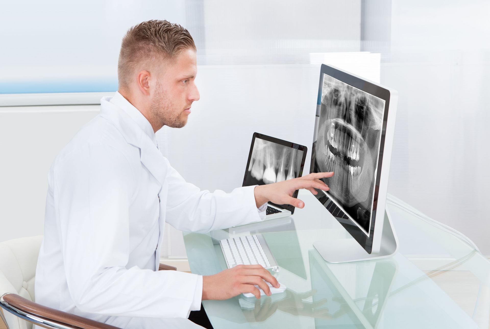
Traditional film X-rays once required considerable time to develop and exposed individuals to higher levels of radiation. Modern digital radiography has completely reshaped this aspect of clinical care by producing instant, high-resolution images that appear on computer monitors within seconds. These digital sensors capture intricate structural details while significantly reducing radiation exposure compared to legacy equipment. Utilizing digital radiography within dental diagnostic services enables practitioners to enlarge images, adjust contrast, and examine hidden tooth structures with high clarity. Early detection of tiny cavities between teeth or subtle changes in bone density allows for prompt, conservative treatment. This technology streamlines office visits, reduces waiting times, and provides individuals with immediate visual feedback regarding their oral health.
Three Dimensional Cone Beam Computed Tomography
Two-dimensional imaging provides valuable information, but certain complex clinical scenarios require a complete three-dimensional view of the oral cavity and surrounding facial structures. Cone Beam Computed Tomography, often referred to as CBCT, captures detailed three-dimensional scans of teeth, jawbones, nerve pathways, and soft tissue networks in a single quick rotation. Incorporating CBCT scans into dental diagnostic services gives clinical teams a thorough understanding of spatial relationships within the jaw before performing complex procedures. According to IBISWorld, there were 178,602 dental businesses in the U.S. in 2024. As modern practices continue to adopt advanced imaging systems, individuals benefit from higher diagnostic accuracy and meticulously planned treatment protocols that minimize post-procedure recovery times.
Intraoral Cameras for Real Time Visualization
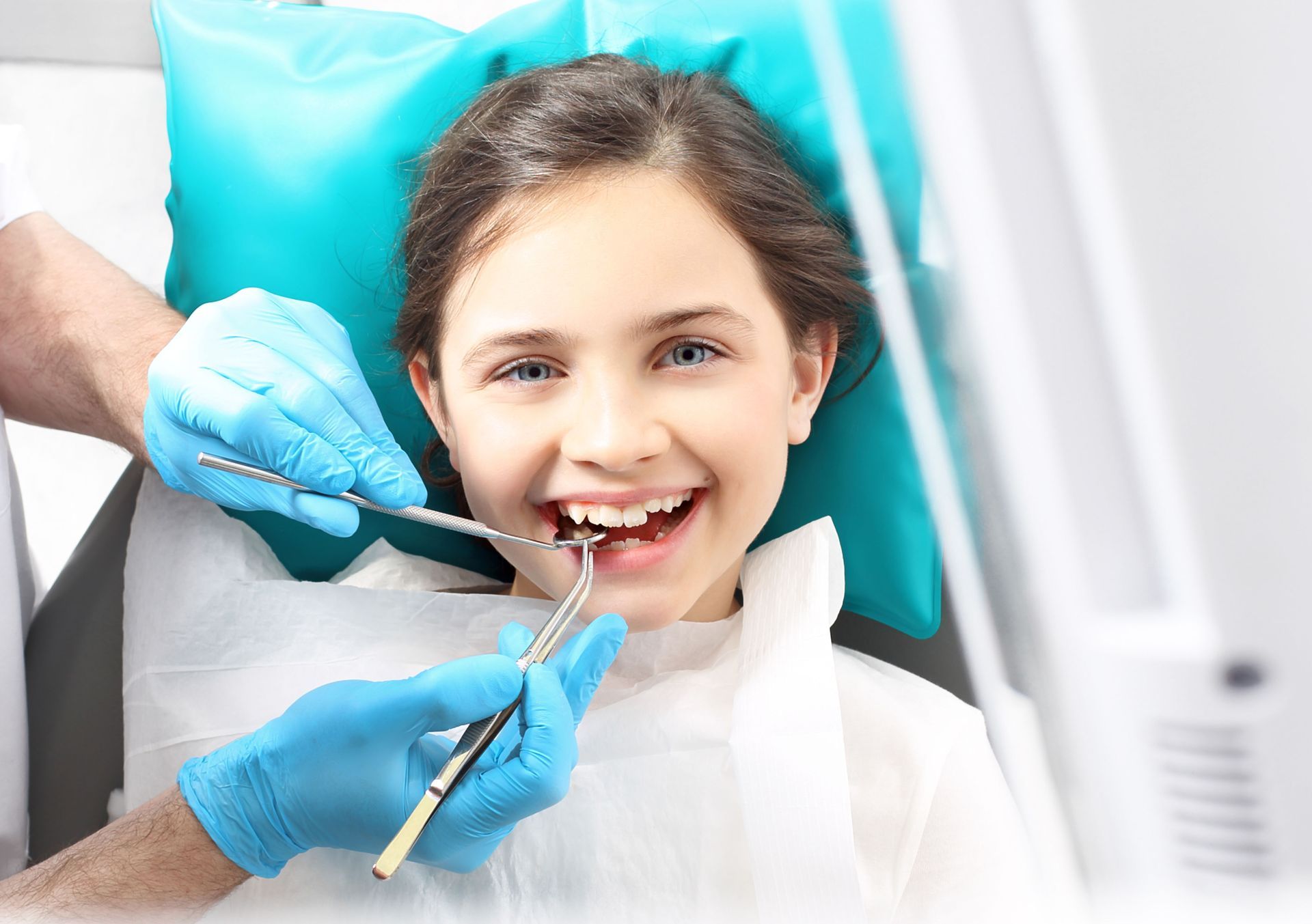
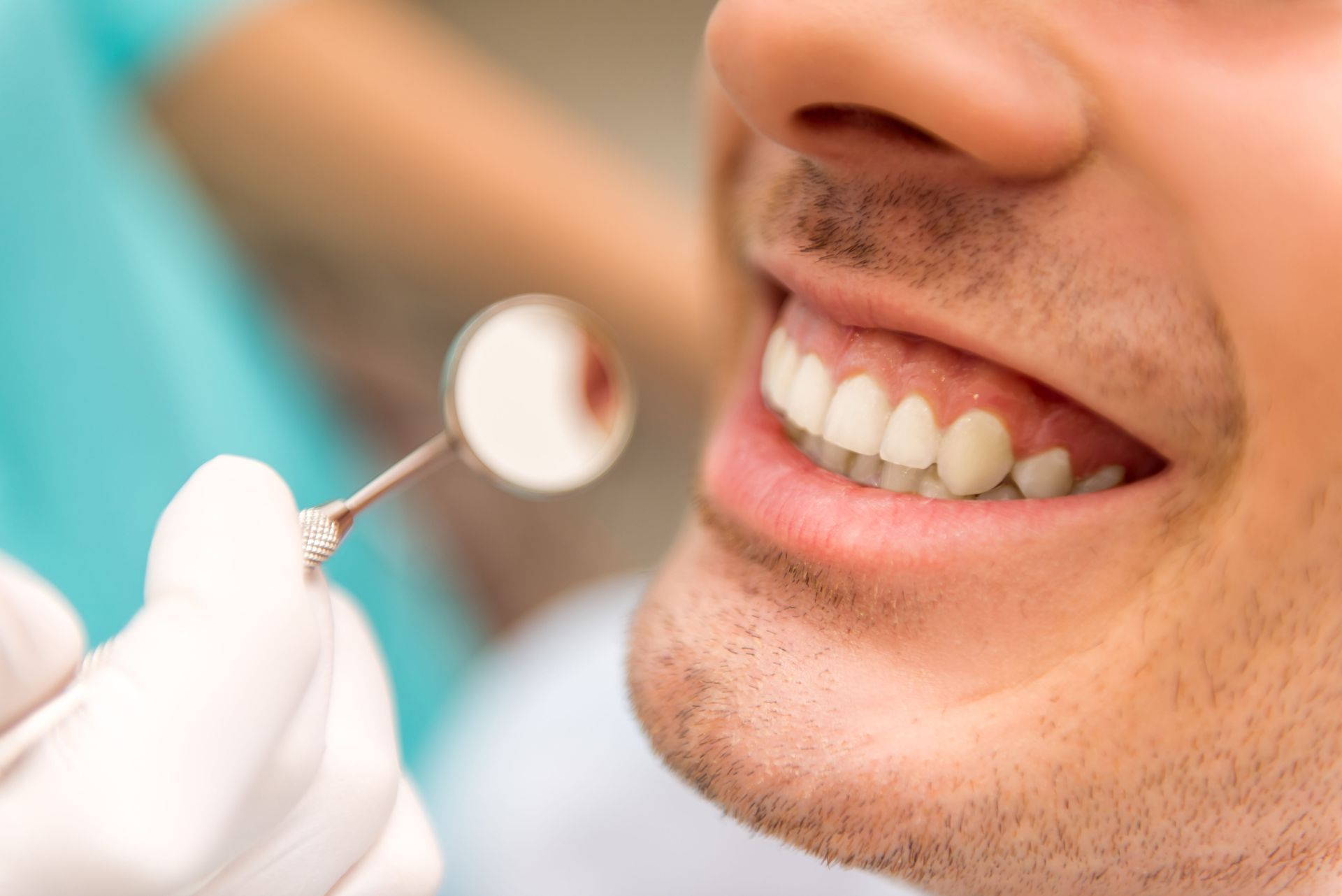
Communicating specific oral health conditions to patients can be challenging when relying solely on verbal descriptions or hand mirrors. Intraoral cameras solve this challenge by using compact, pen-sized digital wands equipped with built-in light sources to capture crisp, magnified images of individual teeth and gum tissue. Featuring these visual tools alongside dental diagnostic services allows individuals to view real-time video footage of their own mouths on chairside monitors. Seeing fractured enamel, worn fillings, or localized gum inflammation firsthand helps patients understand exact clinical findings. This interactive visual feedback encourages meaningful conversations regarding preventive maintenance and proposed care plans, fostering active participation in personal health decisions.
Artificial Intelligence in Radiographic Analysis
Artificial intelligence is rapidly becoming an influential tool across the modern healthcare landscape, particularly within image processing and pattern recognition. Advanced software algorithms can now analyze digital radiographs alongside clinicians, flagging early signs of tooth decay, bone loss, and periapical lesions that might be difficult to spot with the naked eye. Integrating machine learning into dental diagnostic services acts as a reliable second set of analytical eyes, helping standardize diagnostic accuracy across routine examinations. These intelligent algorithms cross-reference subtle image variations against vast databases of clinical data, ensuring consistency and precision. Combining human clinical judgment with algorithmic analysis leads to highly reliable, early-stage diagnoses.
Optical Impression Scanners Replacing Traditional Putty
Taking physical impressions using thick, gooey impression material was long considered one of the most uncomfortable parts of a dental visit. Today, handheld optical scanners capture thousands of precise digital images per second to construct accurate three-dimensional models of the teeth and arches without mess or gagging. Utilizing digital scanners within comprehensive dental diagnostic services streamlines the creation of crowns, aligners, nightguards, and structural bridges. The resulting digital files are transferred instantly to dental laboratories or computer-aided manufacturing units, ensuring exact-fitting restorations. This shift to digital impressions improves physical comfort, reduces appointment lengths, and eliminates the physical waste associated with traditional impression materials.
Laser Fluorescence for Early Cavity Detection
Catching tooth decay at its inception allows for conservative restoration methods that preserve natural enamel structures. Laser fluorescence technology works by emitting specific wavelengths of light onto tooth surfaces, measuring the natural fluorescence returned by healthy versus compromised enamel. Integrating laser detection into routine dental diagnostic services allows clinical teams to evaluate hard-to-reach pits and fissures where decay often begins beneath intact surface enamel. Healthy enamel reflects light differently than decayed tissue, providing a clear numerical reading that indicates the density and health of the tooth structure. Identifying micro-lesions early enables preventive remineralization therapies before physical drilling becomes necessary.
Digital Caries Transillumination Technology
For individuals who require non-ionizing diagnostic methods, near-infrared transillumination offers a brilliant method for detecting interproximal decay and hairline enamel cracks. This technology shines harmless near-infrared light directly through the tooth structure, making the porous enamel appear translucent while areas of decay or structural stress absorb light and appear dark. Using transillumination within dental diagnostic services provides a clear, high-contrast visual representation of tooth integrity without exposing patients to any radiation. It is particularly useful for frequent monitoring of high-risk areas, pregnant patients, or young children. Transillumination complements standard imaging by offering a distinct, radiation-free perspective on enamel health.
Advanced Periodontal Probing Systems
Healthy gum tissue and strong bone support are fundamental to long-term tooth retention, making accurate periodontal evaluation essential. Digital periodontal probes automate the measurement of pocket depths around teeth, recording precise millimeter readings directly into an electronic chart system without manual data entry. Incorporating digital probing tools into routine dental diagnostic services reduces measurement variability and delivers consistent tracking of gum health over time. Automated probing systems apply uniform, gentle pressure during measurements, increasing physical comfort during examinations. The collected data generates detailed graphical charts that highlight areas of inflammation or tissue changes, helping track healing progress after therapy.
Salivary Diagnostics and Biomarker Screening
Saliva contains a wealth of biological information that reflects both oral and systemic health status. Modern laboratory techniques can analyze salivary samples to identify specific bacterial species, viral pathogens, inflammatory proteins, and genetic markers linked to periodontitis and systemic conditions. Including salivary testing alongside comprehensive dental diagnostic services enables personalized risk assessments tailored to an individual's unique biological makeup. Identifying high concentrations of harmful bacteria before physical symptoms manifest allows clinical teams to recommend targeted antimicrobial rinses or specialized home care routines. Salivary diagnostics bridge the gap between oral health and overall physical wellness, supporting proactive healthcare strategies.
Computer Aided Occlusal Analysis
An improper bite can cause unexpected tooth wear, muscle soreness, headaches, and damage to existing dental restorations. Traditional articulating paper shows where teeth touch, but it cannot measure the exact force or timing of those contact points as the jaw moves. Utilizing computerized occlusal analysis within dental diagnostic services provides dynamic, real-time data on how forces are distributed across individual teeth during biting and chewing. Sensors record the exact sequence and intensity of tooth contact, displaying the results through animated visual charts. This precise data allows practitioners to balance bite forces accurately, protecting restorations, relieving jaw tension, and preventing uneven tooth wear.
Technology continues to revolutionize modern oral healthcare, transforming routine examinations into highly accurate, comfortable, and informative experiences. From low-radiation digital imaging and three-dimensional scanning to artificial intelligence and salivary testing, modern diagnostic tools enable early identification of subtle health changes. Early detection allows for gentler, more conservative care plans that protect natural tooth structures and support long-term well-being. Investing in advanced evaluation methods ensures that every patient receives personalized care tailored to their unique needs. To experience advanced diagnostic technology and prioritize your oral health, contact Nurture Dental Health to schedule your next comprehensive examination today.